توضیحات
چکیده
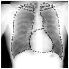
در اين تحقيق مدلي ساده و انعطاف پذير بر مبناي تكنيك مدلهاي توزيع شده نقطه ييا براي بيان تغييرات شكل و ظاهر ريه، قلب و ترقوه در تصاوير راديوگرافي قفسه سينه ارايه شده است. در اين راستا، محدوده مرزي اين اعضا در تصاوير توسط مجموعه ايي از نقاط راهنما مشخص شده و پراكندگي تغييرات از تفاوت نحوهي توزيع اين نقاط راهنما بين ميانگين اشكال و شكل هر عضو در هر تصوير تعيين شده است. روش پيشنهاد شده جهت ساخت مدل، بهينه و داراي پيچيدگي محاسباتي خطي است. اين مدل ميتواند در بسياري از الگوريتم هاي اتوماتيك سگمنت سازي، جستجو و تشخيص سرطان مورد استفاده قرار گيرد. روش ارايه شده بر روي مجموعه ايي شامل 247تصوير راديوگرافي قفسه سينه آزمايش گرديده است. بر اساس نتايج بدست آمده، نود درصد تغييرات فقط توسط تعداد محدودي از پارامترهاي مدل توصيف ميشود. بنابراين يكي از مزاياي استفاده از اين مدل كاهش قابل ملاحظه در حجم محاسبات است .
مقدمه
اندام و اعضاي بدن در بين انسانهـاي مختلـف از نظـر انــدازه ،(Size)شــكل (Shape)و ظــاهر (Appearance)بــا يكديگر تفاوت دارند. بـه همـين علّـت تشـخيص، تحليـل و جداسازيِ اتوماتيك عضو خاصي مثل ريه يا قلب در تصـاوير پزشكي توسط سيسـتمهـاي تشـخيص پزشـكي مبتنـي بـر – بسيار دشوار (Computer- ided Diagnosis) كامپيوتر می باشد. به منظور رفع اين مشكل، تغييرات اندازه، شكل و ظاهر عضو مورد نظر توسط مـدلهـاي مختلفـي بيـان مـيشـود. تاكنون، روش هاي گوناگوني براي ساخت چنين مـدلهـايي ارايه شده است. در مراجع روشهايي كلي به منظـور مدل كردن اين تغييرات ارايه شده است كـه در كاربردهـاي پزشكي نظير اين تحقيق قابل استفاده نميباشند. همچنـين در مراجع روشهاي خاص منظوره مطرح شده اند كـه با وجود كارايي بالا قابل تعميم به كاربردهـاي ديگـر نبـوده، زيرا در اين روشها نياز است تا قالبي (Template) جداگانـه براي هر كاربرد خاص به صورت دستي توسط طـراح تعريـف شود .
ABSTRACT
In this research, a simple and flexible model based on the technique of distributed point-to-point models is presented to express changes in shape and appearance of the lungs, heart and cranial organs in chest radiography images. In this regard, the boundary range of these members in the images is determined by a set of guidance points and the distribution of changes is determined by the difference in distribution of these points between the mean shapes of each member in each image. The proposed method for making the model is optimal and has a linear computational complexity. This model can be used in many algorithms for segmentation, detection, and diagnosis of cancer. The method has been tested on a collection of 247 chest radiography images. Based on the results, ninety percent of the variation is described only by a limited number of model parameters. Therefore, one of the advantages of using this model is a significant reduction in the volume of the calculations.
INTRODUCTION
The organs and organs of the body are different from one another to the size, shape, and appearance of each other. For this reason, the diagnosis, analysis and automatic separation of a particular member, such as the lung or heart, in medical images is based on computer-based diagnostic systems (PCs). In order to solve this problem, changes in the size, shape and appearance of the member are expressed by different models. So far, various methods for making such models have been presented. In the guidelines, general methods have been presented to model these changes that can not be used in medical applications such as this study. Also referred to are general-purpose methods that, despite being high-performance, can not be extended to other applications, because in these methods, a separate template for each specific application needs to be defined manually by the designer.
Year: 2010
Publisher : Eighteenth International Energy Conference of Iran
By : Mohammad Sabri, Alireza Assarat and Bita Shadagar
File Information: persian Language/ 6 Page / size: 863 KB
سال :1389
ناشر : هجدهمین کنفرانس بین المللی برق ایران
کاری از : محمد صبري، عليرضا عصاره و بيتا شادگار
اطلاعات فایل : زبان فارسی / 6 صفحه / حجم : KB 863


نقد و بررسیها
هنوز بررسیای ثبت نشده است.