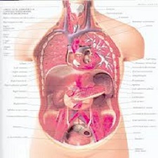
![Radiation.Therapy.for.Liver.Tumors.[taliem.ir]](http://taliem.ir/wp-content/uploads/Radiation.Therapy.for_.Liver_.Tumors.taliem.ir_.jpg)
توضیحات
ABSTRACT
The liver is the central clearing house for most metabolic functions in the body . These functions include lipid, carbohydrate, and protein metabolism; coagulation factor production; albumin production; detoxification of xenobiotics; storage of vitamins and glycogen; and bile processing and secretion. The liver is situated at the receiving end, via the portal circulation, of the intestines, which provide metabolic substrates to the liver. Blood flows out of the liver, carrying away the fruits of its metabolic labor, into the inferior vena cava. Bile flows out of the liver via the bile ducts to aid in digestion and dispose of certain waste products. The liver is for the most part composed of hepatocytes, bile ducts, and blood vessels. Diseases typically target one of these principal components. But, as this is a functional system, injury to one component generally affects other components of the system. The liver has an enormous functional reserve: approximately 80–90% of the liver needs to be destroyed before its essential functions can no longer be adequately performed. Fortunately, the liver is one of the few organs with a high regenerative capacity; this is seen in the ancient Greek story of Prometheus, the giver of fire to humans, who was punished with an endless cycle of having his newly regenerated liver eaten by a bird each day.
INTRODUCTTION
The liver is predominately located in the right upper portion of the abdominal cavity . It normally has a smooth surface contour, is tan-brown in color, and has a weight of 1.4–1.6 kg in an adult. Some of the notable surface landmarks include: from the perspective of the anterior/superior surface, the right lobe and the smaller left lobe of the liver; and from the perspective of the posterior/inferior surface, the quadrate lobe, caudate lobe, gallbladder bed, and porta hepatis (also known as the liver hilum) . Blood flows into the liver through the portal vein and hepatic artery at the porta hepatis. Blood flows out of the liver through the three major hepatic veins, the left, right, and intermediate (middle), at the superior/inferior surface. Bile flows out of the liver through the common hepatic duct at the liver hilum. Anatomic variants exist in the branching and location of blood vessels and bile ducts. The liver can be divided into eight segments based on first and second order divisions of the hepatic artery, portal vein, and bile duct (Fig. 1.1).
چکیده
کبد خانه مرکزی پاکسازی برای اکثر توابع متابولیک در بدن است. این توابع شامل لیپید، کربوهیدرات و متابولیسم پروتئین هستند. تولید فاکتور انعقادی؛ تولید آلبومین؛ سم زدایی از xenobiotics؛ ذخیره سازی ویتامین ها و گلیکوژن؛ و پردازش و ترشح صفراوی. کبد در انتهای دریافت، از طریق گردش پورتال، روده، که مواد زیر ساختی متابولیک را به کبد منتقل می شود. خون از کبد خارج می شود، میوه های کار متابولیک آن را به داخل ورید کبد پایین تر می برد. صفرا از طریق مجاری صفراوی از کبد خارج می شود تا به هضم کمک کند و از مواد زائد خاصی استفاده کند. کبد در اکثر موارد متشکل از hepatocytes، مجاری صفراوی، و عروق خونی است. بیماریها معمولا یکی از این مولفه ها را هدف قرار می دهند. اما، به عنوان این یک سیستم عملکردی، آسیب به یک جزء به طور کلی بر اجزای دیگر سیستم تاثیر می گذارد. کبد دارای ذخایر عملکردی فراوانی است: تقریبا 80-90٪ از کبد باید قبل از اینکه عملکرد های ضروری خود را به اندازه کافی انجام ندهد نابود شود. خوشبختانه، کبد یکی از اندامهای کمی است که توانایی احیاء بالا دارد. این در داستان یونان باستان پرومتئوس دیده می شود که باعث آتش سوزی برای انسان ها شده است، که با یک چرخه بی پایان از داشتن کبد تازه بازسازی شده توسط یک پرنده هر روز خورده می شود.
مقدمه
کبد عمدتا در قسمت فوقانی سمت راست حفره شکمی واقع شده است. به طور معمول دارای یک سطح سطح صاف است، قهوه ای مایل به قهوه ای است و دارای وزن 1.4-1.6 کیلوگرم در بزرگسالان است. برخی از نشانه های سطح قابل توجه عبارتند از: از منظر سطح قدام / برتر، لوب راست و لوب کبد کوچکتر کبد؛ و از منظر سطح خلفی / پایین تر، لوب کوادرات، لوب کادت، بستر کیسه صفرا، و هپاتیس پرتا (همچنین به عنوان گوسفند کبد شناخته می شود). خون به داخل کبد از طریق ورید پورتال و سرخرگ کبدی در خون انتقال می یابد. خون در خارج از کبد از طریق سه رگهای کبدی بزرگ، چپ، راست و متوسط (وسط) در سطح بالاتر / پایین تر جریان می یابد. صفرا خارج از کبد از طریق مجرای کبدی معمولی در کبد جریان دارد. انواع آناتومی در شاخه و محل رگ های خونی و مجاری صفرا وجود دارد. کبد را می توان به هشت بخش بر اساس بخش اول و دوم مرتبه کبد، ورید پورتال و مجرای صفرا تقسیم کرد (شکل 1.1).
Year: 2016
Publisher: SPRINGER
By : Jeffrey Meyer, Tracey E. Schefter
File Information: English Language/ 285 Page / size: 5.90 MB
Only site members can download free of charge after registering and adding to the cart
سال : 1395
ناشر : SPRINGER
کاری از : جفری میر، تراسی اچ. چیپتر
اطلاعات فایل : زبان انگلیسی / 285 صفحه / حجم : MB 5.90
![A double-blind controlled study of a nonhydroquinone bleaching[taliem.ir]](http://taliem.ir/wp-content/uploads/A-double-blind-controlled-study-of-a-nonhydroquinone-bleachingtaliem.ir_.jpg)
![Development of prebiotic food products and health benefits[taliem.ir]c](http://taliem.ir/wp-content/uploads/Development-of-prebiotic-food-products-and-health-benefitstaliem.ir_.jpg)

![Organizing.Patient.Safety.Failsafe.Fantasies.[taliem.ir]](http://taliem.ir/wp-content/uploads/Organizing.Patient.Safety.Failsafe.Fantasies.taliem.ir_-150x150.jpg)
![Recurrent.Respiratory.Papillomatosis.[taliem.ir]](http://taliem.ir/wp-content/uploads/Recurrent.Respiratory.Papillomatosis.taliem.ir_-150x150.jpg)
نقد و بررسیها
هنوز بررسیای ثبت نشده است.